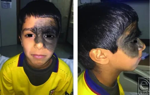
What Are Congenital Nevi?
Congenital nevi are pigmented birthmarks that are present at birth or appear within the first few weeks of life. These lesions develop from melanocytes, the pigment-producing cells in the skin, which cluster together during fetal development instead of spreading evenly. Plastic surgery congenital nevi procedures have become increasingly refined, offering families effective solutions for managing these lesions both medically and cosmetically.
Congenital melanocytic nevi (CMN) occur in roughly 1% of all newborns, making them one of the most common types of birthmarks encountered in pediatric practice. While the vast majority of these lesions are small and benign, larger nevi can raise significant medical concerns, including an elevated risk of melanoma and potential psychosocial challenges as the child grows.
The formation of congenital nevi occurs during the first trimester of pregnancy, typically between the fifth and twenty-fourth week of gestation. These lesions result from localized genetic abnormalities—most commonly activating mutations in the NRAS gene within the MAPK signaling pathway—that cause melanocytes to over-proliferate in specific areas. There is currently no known way to prevent a congenital nevus from forming.
Classification of Congenital Melanocytic Nevi
Understanding the classification of congenital nevi is essential because the size of the lesion directly influences treatment decisions, melanoma risk, and surgical planning. Congenital melanocytic nevi are categorized based on their projected adult size (PAS), which accounts for proportional growth as the child matures.
Size-Based Classification
| Category | Diameter | Incidence | Melanoma Risk |
| Small | < 1.5 cm | Very common | Very low |
| Medium | 1.5 – 19.9 cm | Common | Low to moderate |
| Large | 20 – 40 cm | Rare | Elevated (1–2%) |
| Giant | > 40 cm | ~1 in 20,000 | Significant (5–15%) |
The scaling factor used to predict adult size varies by anatomical location. A congenital nevus on the head is expected to grow by a factor of approximately 1.7, while those on the lower limbs may grow by a factor of 3.3, and lesions on the upper limbs and torso by roughly 2.8. This projection helps plastic surgeons plan the scope and timing of surgical intervention.
A more refined classification system further subdivides medium nevi into M1 (1.5–10 cm) and M2 (10–20 cm), large nevi into L1 (20–30 cm) and L2 (30–40 cm), and giant nevi into G1 (40–60 cm) and G2 (greater than 60 cm). When more than 50 satellite lesions are present, the size category is increased by one level to account for the greater total nevus burden and associated melanoma risk.
Clinical Features and Symptoms
Congenital nevi are often visible at birth, initially appearing as flat, pale-brown patches. Over time, these lesions can undergo several changes that may prompt medical evaluation and referral to a plastic surgeon.
Common Characteristics
Pigmentation changes: Congenital nevi may lighten or darken over time. Some lesions develop uneven pigmentation, which can make clinical monitoring more challenging.
Texture changes: Initially flat lesions may become raised, develop a verrucous or pebbly surface, or form proliferative nodules. These changes are common and do not necessarily indicate malignancy, though they warrant professional evaluation.
Hypertrichosis: Excess hair growth over the nevus is common, particularly in larger lesions. Coarse, dark hair typically appears when the child is one to two years of age, which is why giant congenital nevi are sometimes called giant hairy nevi.
Satellite lesions: Smaller pigmented spots scattered around the primary nevus are known as satellite nevi. Their presence and number are clinically significant and factor into risk assessment and treatment planning.
Associated Conditions
Neurocutaneous melanocytosis (NCM): Large and giant congenital nevi can grow internally, potentially affecting the central nervous system. NCM involves the deposition of melanocytes within the brain and spinal cord and can be associated with seizures, increased intracranial pressure, headaches, and visual disturbances. Screening MRI of the central nervous system in the first year of life is recommended for children with large or giant CMN, as an abnormal MRI has been identified as the strongest predictor of melanoma risk across all sites.
Why Plastic Surgery for Congenital Nevi?
The decision to pursue plastic surgery congenital nevi treatment involves careful consideration of multiple factors. Plastic surgeons bring specialized expertise in both removing the lesion and reconstructing the affected area with optimal aesthetic results.
Medical Reasons for Surgical Removal
Melanoma risk reduction: While the effectiveness of surgical excision in completely eliminating melanoma risk remains debated—since melanoma can occasionally develop even after removal—excision does reduce the total number of potentially malignant melanocytes. For large and giant nevi with lifetime melanoma risks estimated between 5% and 15%, this risk reduction is clinically meaningful.
Functional considerations: Nevi located near the eyes, nose, joints, or other critical areas may interfere with normal movement, development, or function. Early removal can prevent these functional limitations.
Reduced monitoring burden: Complete removal of a congenital nevus eliminates the need for lifelong surveillance of that particular lesion, reducing anxiety for both the patient and family.
Psychosocial and Cosmetic Reasons
Recent research has shown that treatment indications for congenital nevi have shifted increasingly from prevention of malignant transformation toward improvement of appearance and psychosocial health. Many families choose removal to help their child avoid the self-esteem issues and unwanted attention that visible birthmarks can cause, particularly as children enter school age. Board-certified plastic surgeons are uniquely qualified to achieve the best aesthetic outcomes while addressing the underlying medical concerns.
Plastic Surgery Techniques for Congenital Nevi Removal
The surgical approach to congenital nevi removal depends on the size, location, and depth of the lesion, as well as the patient’s age and overall health. Plastic surgeons employ several techniques, often in combination, to achieve complete removal with the best possible cosmetic result.
Simple Excision
For small and some medium-sized congenital nevi, simple excision remains the most straightforward approach. The surgeon removes the entire lesion along with a small margin of surrounding healthy tissue and closes the wound with sutures. This technique is most effective when there is sufficient surrounding skin laxity to allow tension-free closure. In many cases, absorbable sutures can be used, eliminating the need for a separate appointment for suture removal.
Serial Excision
When a nevus is too large to remove in a single procedure, serial excision is performed. This technique involves removing a portion of the lesion during each surgery, with the wound closed after each stage. Over multiple procedures spaced weeks to months apart, the entire nevus is gradually removed. Between surgeries, the surrounding skin naturally softens and stretches, allowing progressively larger portions to be removed without placing excessive tension on the closure. Serial excision is particularly effective for medium to large nevi that are not amenable to single-stage removal.
Tissue Expansion
Tissue expansion is one of the most effective techniques for treating large and giant congenital nevi, often producing the best cosmetic outcomes in terms of both appearance and function. The procedure involves placing one or more silicone balloons (expanders) beneath the healthy skin adjacent to the nevus. Over a period of several weeks, the expanders are gradually inflated with saline through a small injection port, slowly stretching the overlying skin.
Once sufficient extra skin has been generated, the expanders are removed, the nevus is excised, and the newly expanded skin is advanced to cover the defect. Because the expanded skin matches the color, texture, and hair-bearing characteristics of the surrounding area, the aesthetic results are typically superior to those achieved with skin grafting.
Skin Grafting
Skin grafting involves removing the nevus and covering the wound with skin harvested from another area of the patient’s body (the donor site). Full-thickness skin grafts (FTSG) provide better color match and texture compared to split-thickness grafts, making them preferable for visible areas such as the face. However, skin grafting may leave noticeable differences in pigmentation between the grafted and surrounding skin, and additional cosmetic treatments may be required later. This technique is often used when tissue expansion is not feasible due to the location or extent of the nevus.
Curettage and Dermabrasion
Curettage involves scraping away the superficial layers of the nevus, while dermabrasion uses specialized instruments to mechanically remove the outer layers of skin. These procedures are most effective when performed during the first few months of life, when the boundary between the nevus and underlying tissue is most distinct. They are primarily used for lesions on the head and face where surgical excision may be particularly challenging.
However, these superficial techniques carry a significant risk of color recurrence and do not remove deeper nevus cells. Additional treatments, including laser therapy and further surgical excision, are frequently required after the initial procedure.
Laser Therapy
Several laser modalities can be used as adjunctive treatments for congenital nevi, including Q-switched ruby lasers, Nd:YAG lasers, and CO2 lasers. Laser therapy is most commonly used to address residual pigmentation after curettage or to treat areas where complete surgical excision is not possible. While laser treatment can improve the cosmetic appearance of nevi, it does not eliminate melanoma risk because nevus cells in the deeper dermal layers remain intact. Multiple treatment sessions are typically required.
Optimal Timing for Surgery
The timing of plastic surgery congenital nevi procedures is an important consideration that varies based on individual circumstances.
Early Intervention
Many pediatric plastic surgeons recommend beginning treatment as early as six months of age for several reasons. Infant skin is more pliable and heals with less scarring than older skin. Early removal means the child’s nevi are treated while the lesions are smaller in absolute terms, potentially simplifying the surgical approach. Additionally, completing treatment before the child enters school helps avoid the psychosocial impact of visible birthmarks during formative years.
For curettage procedures specifically, the first few months of life represent the optimal window, as the nevus cells are more superficially located and easier to separate from the underlying tissue at this stage.
Staged Treatment Planning
For large and giant congenital nevi, treatment is typically staged over several years. The goal is to complete the majority of surgical procedures before the child begins school. A comprehensive treatment plan may combine multiple techniques—such as tissue expansion followed by serial excision and skin grafting—performed sequentially to achieve the best possible outcome.
Risks and Potential Complications
As with any surgical procedure, congenital nevi removal carries certain risks that patients and families should understand before proceeding.
Surgical Risks
- Scarring: All surgical removal will result in some degree of scarring. Plastic surgeons employ specialized techniques to minimize scar visibility and optimize healing.
- Infection: As with any surgery, there is a risk of wound infection, which can delay healing and reconstruction.
- Pigmentation differences: Grafted skin or expanded skin may not perfectly match the surrounding area in color or texture, potentially requiring future cosmetic treatments.
- Anesthesia risks: Most congenital nevi removals in children require general anesthesia, which carries its own set of risks that should be discussed with the anesthesiology team.
- Recurrence: Some nevus cells may remain after treatment, particularly with superficial techniques like curettage and laser therapy, leading to color recurrence or regrowth.
Important Considerations About Melanoma Risk
It is important to understand that surgical removal of a congenital nevus does not completely eliminate the risk of melanoma. Melanoma has been reported even after complete surgical excision, and approximately one-third of melanomas in patients with CMN may involve the central nervous system due to neurocutaneous melanocytosis. Scarring from removal can also potentially mask the development of melanoma, complicating future detection. For these reasons, long-term dermatological follow-up is recommended for all patients, regardless of whether the nevus has been surgically removed.
What to Expect: Before, During, and After Surgery
Pre-Surgical Consultation
During the initial consultation, a board-certified plastic surgeon will evaluate the nevus, discuss the family’s concerns and goals, and develop a comprehensive treatment plan. This evaluation typically includes a thorough physical examination, documentation of the nevus with photographs, and possibly a referral to dermatology for dermoscopic evaluation or biopsy if there are any concerning features. For children with large or giant nevi, an MRI of the central nervous system may be recommended to screen for neurocutaneous melanocytosis.
The Surgical Experience
Most congenital nevi surgeries in children are performed under general anesthesia. The specific procedure and its duration will depend on the technique being used and the extent of the nevus. Smaller excisions may be performed on an outpatient basis, while more complex procedures involving tissue expansion or extensive reconstruction may require overnight hospitalization for monitoring.
Recovery and Aftercare
Recovery varies depending on the type of procedure performed. For simple excisions, children typically heal within two to three weeks with proper wound care. Tissue expansion procedures involve a longer process, with the expansion phase lasting several weeks before the final reconstruction. Dark patches of skin may appear around surgical scars, which can sometimes be addressed with additional treatment if needed.
Many patients with large or giant congenital nevi will require lifelong outpatient follow-up care after completing their surgical treatment, but most face few functional limitations once fully healed.
Choosing the Right Plastic Surgeon
Selecting the right surgeon for plastic surgery congenital nevi treatment is one of the most important decisions families will make. Key qualifications to look for include:
- Board certification in plastic and reconstructive surgery
- Fellowship training or specialized experience in pediatric plastic surgery
- Demonstrated experience with congenital nevi removal and reconstruction
- A compassionate, family-centered approach to care
- Access to a multidisciplinary team including dermatologists, pediatricians, and anesthesiologists experienced with pediatric patients
An interprofessional team approach is strongly recommended for managing children with congenital melanocytic nevi, particularly for larger lesions. This team typically includes pediatricians, dermatologists, plastic surgeons, and when necessary, neurologists and oncologists.
Frequently Asked Questions About Plastic Surgery Congenital Nevi
Are all congenital nevi dangerous?
No. Most small and medium-sized congenital nevi are benign and pose only a slightly elevated risk of melanoma compared to normal skin. However, large and giant nevi carry a more significant risk and require careful monitoring and often surgical intervention.
At what age should my child have surgery?
The optimal timing depends on the size, location, and characteristics of the nevus. Many surgeons recommend beginning treatment as early as six months of age, with the goal of completing major procedures before the child starts school. Your plastic surgeon will develop a personalized timeline based on your child’s specific situation.
Will the nevus come back after surgery?
Complete surgical excision is typically curative for the removed area. However, superficial techniques like curettage and laser therapy carry a higher risk of recurrence, and satellite lesions may still develop independently. Ongoing monitoring is recommended regardless of the treatment method used.
How many surgeries will my child need?
The number of procedures depends on the size and complexity of the nevus. Small nevi may be removed in a single surgery, while large or giant nevi may require multiple staged procedures over several years, combining techniques such as tissue expansion, serial excision, and skin grafting.
Will insurance cover the surgery?
Many insurance plans cover congenital nevi removal when there is a documented medical indication, such as elevated melanoma risk or functional impairment. Coverage may vary for procedures performed primarily for cosmetic reasons. It is advisable to consult with both your surgeon’s office and your insurance provider to understand your specific coverage.
Does surgical removal completely eliminate the risk of melanoma?
No. While removing the nevus reduces the number of potentially malignant cells, melanoma can occasionally develop even after complete excision. Additionally, patients with large or giant nevi remain at risk for central nervous system melanoma. Lifelong follow-up with a dermatologist is recommended for all patients with a history of congenital melanocytic nevi.
Conclusion
Plastic surgery congenital nevi treatment represents a vital intersection of reconstructive expertise, oncological awareness, and compassionate pediatric care. Advances in surgical techniques—from tissue expansion to refined serial excision methods—have made it possible to achieve remarkable aesthetic outcomes while addressing the underlying medical risks associated with these birthmarks.
The decision to pursue surgery is deeply personal and should be made collaboratively between families and a qualified multidisciplinary medical team. Whether the primary motivation is reducing melanoma risk, improving cosmetic appearance, or supporting a child’s psychosocial well-being, today’s plastic surgeons have the tools and expertise to deliver safe, effective, and life-changing results.
If your child has a congenital nevus, we encourage you to schedule a consultation with a board-certified plastic surgeon who specializes in pediatric reconstructive procedures. Early evaluation and proactive planning can make a significant difference in outcomes and provide families with the peace of mind they deserve.